Necessity is the Mother of Invention

Jefferson Docs Find New Uses for Existing Tools
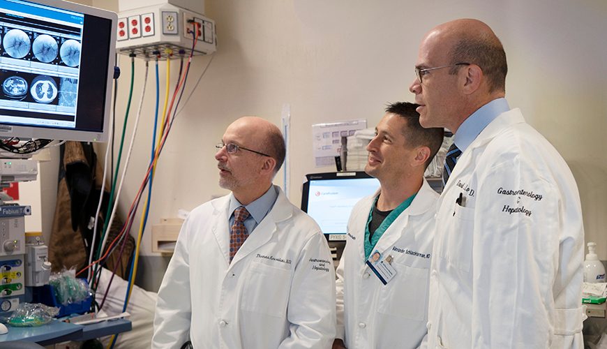
Some doctors look at a piece of medical equipment and appreciate what can be done with it. Thomas E. Kowalski, MD, and David E. Loren, MD, look at that same piece of equipment and appreciate what else can be done with it.
Thinking outside the box and coming up with inventive ways to use existing tools to best benefit patients are these gastroenterologists’ stock-in-trade. Kowalski and Loren are two of the six physicians who make up the pancreaticobiliary and advanced endoscopy section of Thomas Jefferson University Hospital’s Division of Gastroenterology and Hepatology.
“The types of things that we do here are pushing the boundaries of innovation along many different fronts,” Loren says. “Our unique and cutting-edge endoscopic procedures are changing the face of medicine and will become the standard of care—and we’re at the forefront of it.”
The pancreaticobiliary and advanced endoscopy section specializes in advanced endoscopic procedures and technologies. Loren explains it’s not the state-of-the-art technology that sets them apart—it’s the creative and unusual ways in which they use it.
The basis of their work uses the endoscope, a long, thin, flexible tube that contains high-resolution optical sensors. These sensors transmit images and provide working channels through which devices can be passed to the inner reaches of the gastrointestinal tract, including the pancreas and bile duct. New instruments include cutting and suturing devices that allow doctors to perform repairs and resections without making skin incisions.
For example, the doctors can now close fistulas (abnormal openings connecting two parts in the body) and remove tumors in the gastrointestinal tract. In the past, someone suffering from achalasia—a disorder where the muscle at the bottom of the esophagus is too tight to let food pass—would need complicated surgery; endoscopy allows doctors to repair the condition by cutting the muscle from the inside of the esophagus in a same-day procedure.
These endoscopic methods are noninvasive and performed on an outpatient basis, resulting in fewer complications, shorter treatment time, and faster recovery.
“Things that used to be done by surgery—both for diagnosis and for treatment—can now be done without making a cut,” Kowalski says.
By pairing endoscopy with other tools, Loren says the sky’s the limit.
“We have endoscopic ultrasound, radiology, laser, steerable wires, needles to retrieve and deliver, probes that deliver heat or cold,” he says. “We think about what the patient needs and say to ourselves, ‘OK, what tools are available, and how can I creatively use all the things at my disposal to come out with the least invasive and best outcome for this individual?’”
To Kowalski, necessity is the mother of invention, and the “innovative thought process” is what leads to novel uses. “We’ll be in a procedure and say, ‘If we only had X, we could do Y.’ Then we brainstorm together, modify this or that and often make Y happen, but also document how it could be done even better.”
One of their most useful and versatile tools is the humble stent. Approximately 20 millimeters by one centimeter and made of a titanium-nickel alloy with a silicone coating, the tube-shaped device can, in many instances, replace the need for invasive surgery.
Kowalski ticks off just a few of the applications that help to avoid surgery: “If a patient has a cancer that is blocking the stomach and preventing the patient from eating, we can use endoscopy to place a stent to create a pathway for food. In the case of a patient suffering from inflammation of the gall bladder, but is too sick to undergo surgery to have the organ removed, we can place a stent to drain the gall bladder internally into the intestine. For those who have undergone gastric bypass and develop complications with their bile ducts, we can use the stent to access the bypassed stomach and the nearby bile duct, rather than perform a complex surgery.”
Novel uses of endoscopic ultrasound are also bringing cancer care to new levels, as the technology enables doctors to perform procedures outside of the gastrointestinal tract, where traditional endoscopy is limited. The technique allows for directed treatments for certain malignancies. Only a handful of academic research hospitals across the country are doing this, and Jefferson has been consistently at the forefront of innovative ways of using endoscopy. The team has collaborated with industry developers to participate in studies that have led to FDA approval for devices and procedures.
Kowalski and Loren both admit they cannot “leave well enough alone.” They recently began a study into the use of contrast-enhanced endoscopic ultrasound for esophageal cancer to determine whether cancer is present in the primary draining lymph node. The test would indicate the likelihood that the cancer has spread to other parts of the body. “Contrast agents are injected into a tumor to follow the path of the contrast agent and determine what lymph node to sample,” Loren says. “The study will allow us to map how and where tumors spread. This combines endoscopic technology, ultrasound technology, and microbubble contrast technology to determine how we can better care for patients.”
Both doctors relish the advances the next 10 years will bring, but stress that innovations in medical care depend heavily on research funding.
“These revolutionary advances come from the minds of the physicians who embrace clinical investigation in order to meet the needs of patients,” Loren says. What begins as a “what if” turns into “let’s try this.” In the beginning, pilot programs are unfunded and depend on the volunteer time of the doctors, fellows, and staff members. Once enough data is collected, the research group seeks corporate, organizational, and/or private funding.
Kowalski emphasizes the importance: “Only through philanthropy can we move technology forward and find ways of changing the trajectory of medicine.”